Extracellular vesicles are a series of membrane-structured vesicles released by cells, including exosomes, microvesicles, and apoptotic bodies. Extracellular vesicles are composed of various lipids and membrane proteins, some of which special membrane proteins help target specific tissues and cells, while others ensure that non-specific interactions are minimized, these physiological properties endow Thanks to their excellent biocompatibility and ability to target lesions, extracellular vesicles can be widely used in research in the field of drug delivery.
Compared with exosomes and microvesicles produced by living cells, apoptotic bodies derived from apoptotic cells have a higher production efficiency. Apoptosis is a natural process in which cells actively package biomolecules into vesicles. Based on this, drugs can be efficiently loaded into apoptotic bodies through this process, so the complete apoptotic body can be used as a potential drug delivery vehicle. Some apoptosome derivatives (such as recombinant apoptosomes, apoptosome membrane-modified nanoparticles, etc.) not only retain the excellent physiological properties of apoptosomes, but also have the appropriate size required for drug delivery and higher the drug loading efficiency, which can further promote the application of apoptosome-based drug delivery system.
Mechanism of Apoptotic Body Formation

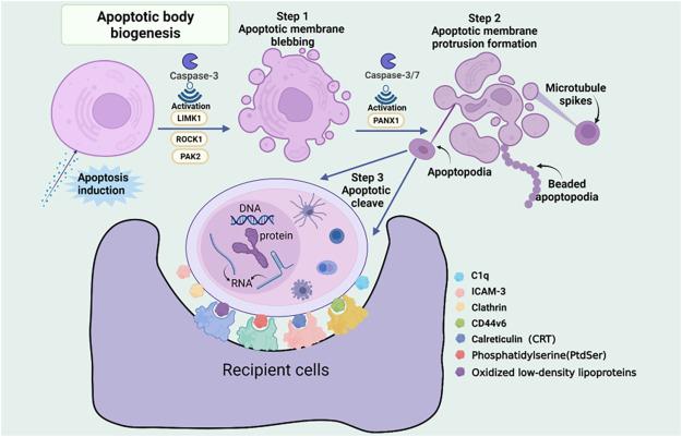
Apoptosis is a conserved form of programmed cell death that usually occurs in multicellular organisms to maintain the homeostasis of normal cell populations in the organism and thus plays a crucial role in development and aging. This programmed cell death process is mediated by related signaling pathways triggered by multiple factors, including cellular stress, DNA damage, and immune surveillance. Throughout the process of apoptosis, cells undergo a series of morphological changes that eventually lead to the breakdown of apoptotic cells. The disassembly of apoptotic cells is divided into three distinct morphological processes, namely, plasma membrane blebbing, formation of apoptotic membrane protrusions, and final resolution into apoptotic bodies.
- Plasma membrane blebbing
Plasma membrane blebbing is the first step in the disassembly of apoptotic cells, that is, the formation of large circular bumps on the surface of apoptotic cells, which is driven by the collapse of the cytoskeleton and increased hydrostatic pressure, while the compressive stress generated during actomyosin contraction increases the hydrostatic pressure inside the cell, allowing cytosol to squeeze out from areas of weak membrane attachment, ultimately leading to plasma membrane blebbing. This process results in the formation of apoptotic bodies that retain abundant cellular contents and associated protein molecules. At the molecular level, the plasma membrane blebbing process is regulated by a variety of kinases, including p21-activated kinase 2 (PAK2), Lim domain kinase 1 (LIMK1) and Rho-associated kinase 1 (ROCK1), among which ROCK1 is considered to be the A key positive regulator of the membrane blebbing process.
- Formation of Apoptotic Membrane Protrusions
After plasma membrane blebbing, some cells can continue to produce long apoptotic membrane protrusions, called apoptopodia and beaded apoptopodia. Apoptotic feet are extruded from the plasma membrane after blebbing and have a long string-like appearance. Apoptotic body-like structures can be observed along these protrusions. It can be seen that apoptotic feet promote apoptosis body formation by separating membrane vesicles. In contrast, the beaded apoptotic podia are connected by several continuous apoptotic bodies, and their length can reach several times that of the parental cell, and the breakage of a single beaded apoptotic podia can result in 10-20 apoptotic bodies of the same size. Apoptotic bodies are released, and the diameter of each apoptotic body is 1-3 μm.
- Apoptotic Body Formation
Fragmentation of apoptotic membrane protrusions to form dispersed apoptotic bodies is the last step in the disassembly of apoptotic cells. Usually this process requires the cooperation of external factors (such as shear force, physical interaction between cells) and internal factors (such as shedding-like processes). Many factors that regulate upstream steps of cellular disassembly affect the production of apoptotic bodies. For example, inhibition of ROCK1, actin polymerization, and vesicle trafficking not only inhibits plasma membrane blebbing or apoptotic membrane protrusion formation, but also leads to a reduction in the overall production of apoptotic bodies. In contrast, inhibition of PANX1 activity enhanced the formation of apoptotic membrane protrusions, resulting in a subsequent increase in the number of apoptotic bodies. In addition, cells lacking LIMK1 produced more apoptotic bodies after induction of apoptosis. Based on the current understanding of the mechanism of the decomposition process of apoptotic cells, the formation of apoptotic bodies is considered to be a process of biological development that can be intervened.
Classes of Apoptosome-Based Drug Delivery Systems
Apoptotic bodies can load a series of cellular contents (such as DNA, RNA, and protein, etc.), and transfer these substances to other cells that can phagocytose apoptotic bodies, promoting the exchange of materials and information between cells, which indicates that apoptotic bodies have great potential as drug delivery vehicles. At present, drug delivery systems based on apoptotic bodies are mainly divided into four categories:
- Complete Apoptotic Bodies Directly Used as Drug Carriers
Various stress methods are used to induce apoptosis, and the resulting apoptotic bodies are collected to construct drug-loaded apoptotic bodies. These drug-loaded apoptotic bodies retain the relevant biological characteristics of the parental cells, showing excellent ability to target the lesion.
- Recombinant Apoptotic Body Drug Delivery Systems
In order to retain the biocompatibility and targeting ability of the apoptotic body, and improve the efficiency and stability of drug loading, the researchers first mixed the prepared apoptotic body with the drug, and then reorganized the apoptotic body through various mechanical effects.
- Drug Delivery Based on In Situ Generation of Apoptotic Bodies System
After apoptosis produces apoptotic bodies, they can not only be engulfed by professional phagocytes, but also can be engulfed and processed by neighboring cells. Based on this neighborhood effect, the apoptotic body can carry the remaining drug to adjacent tumor cells after apoptosis to realize the deep penetration of the drug in the tumor.
- Apoptotic Body-Like Biomimetic Delivery System Delivery System
Inspired by the ability of phagocytes to specifically phagocytose apoptotic bodies, researchers designed a series of apoptosome-like biomimetic nano-drug carriers to achieve drug delivery to macrophages. These biomimetic nano-drug carriers not only retain the excellent targeting ability of apoptotic bodies, but also have the advantages of easy preparation and modification of nano-drug carriers.